

The world of cardiac imaging is moving at an incredible pace. What started as simple chest scans has become a high-tech reality. Today, we have advanced systems that show the heart in motion, in great detail, and in real time. Doctors are no longer just looking at flat, still pictures. They are now analyzing exactly how the heart beats and responds to stress. This massive shift is changing the way we look at healthcare.
From the old-school echocardiogram test to the modern wonder of 4D ultrasound, technology is getting faster and smarter. These advancements help doctors find heart problems much earlier than before. They can plan surgeries more effectively and help patients recover faster. In this guide, you will see how cardiovascular imaging has changed. We will look at the trends shaping the Future of cardiac imaging and why 4D cardiac imaging is the next big thing.
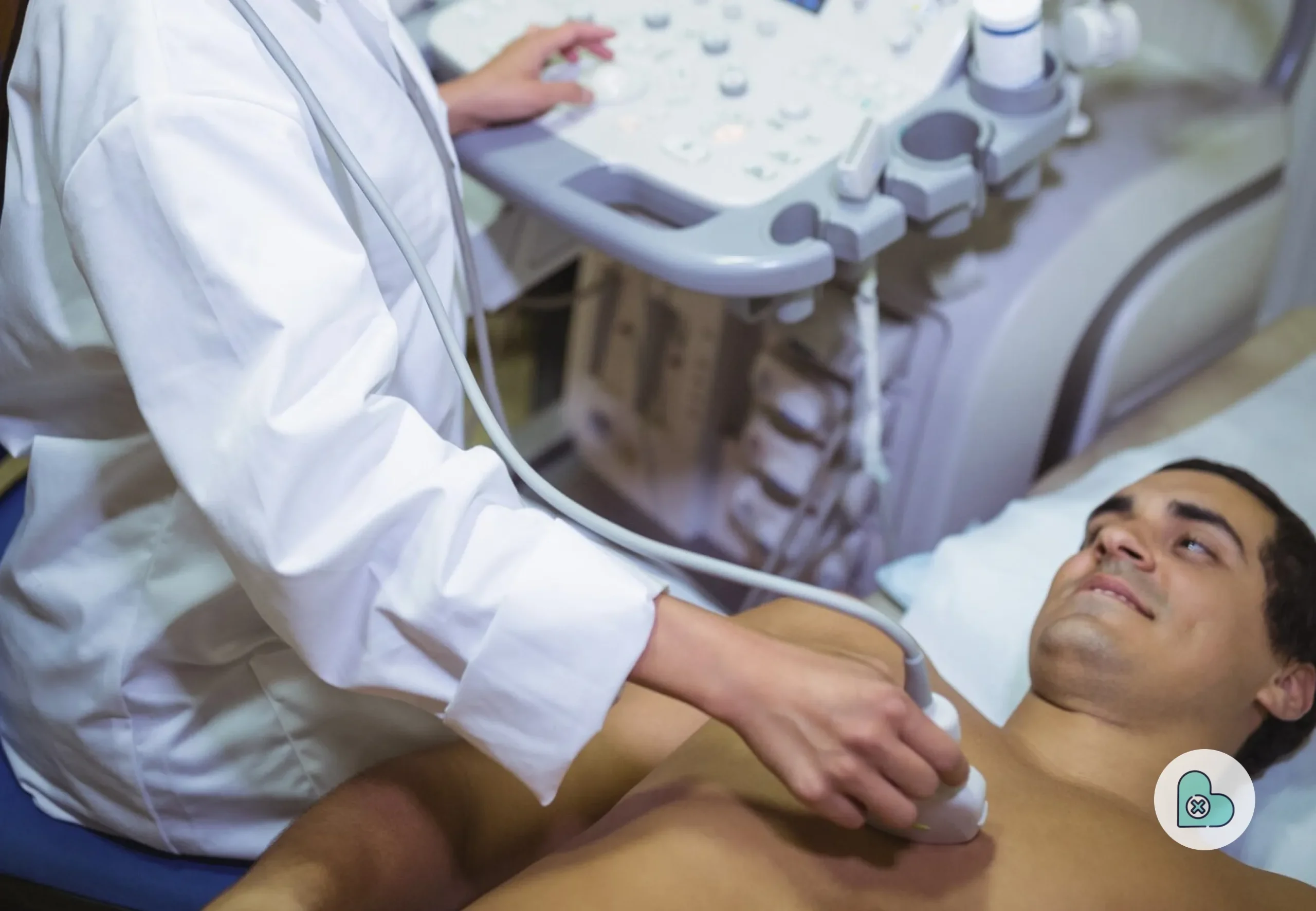
What Exactly is Cardiac Imaging?
_ iStock
Cardiac imaging is a group of advanced techniques used to visualize the heart without surgery. It is a completely non-invasive cardiac imaging process that keeps the patient safe. Doctors use these tools to understand the size and shape of the heart. They can check how well the heart pumps blood and look for leaks in the heart valves. Since heart disease is a leading killer worldwide, having clear heart scan technology is vital.
With the right advanced cardiac imaging, doctors can detect diseases before they become emergencies. They can monitor ongoing issues and plan treatments that actually work. In simple terms, this technology gives a clear window into the chest. It helps the medical team see exactly what is happening inside. This leads to better heart disease prevention and helps keep people healthier for longer.
The Evolution of Cardiac Imaging Technology
_ istockphoto
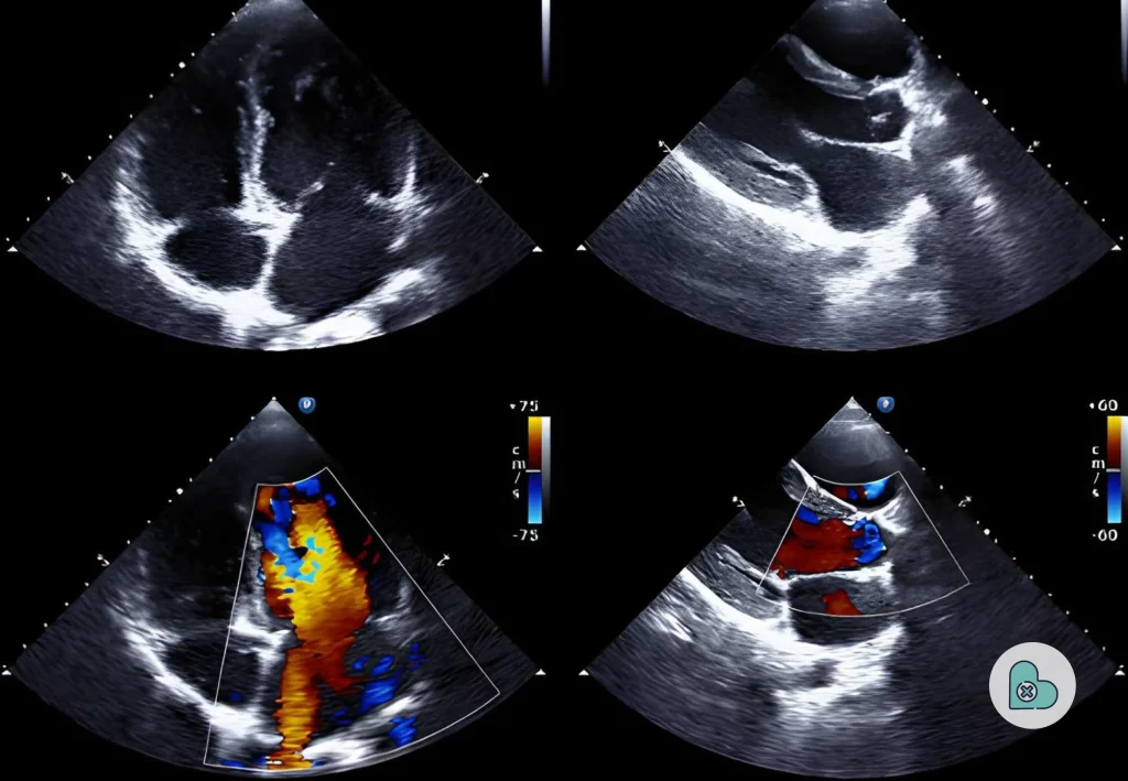
1. Traditional Echocardiography
For many years, echocardiography has been the main tool for heart doctors. It uses sound waves to create live images of the heart. This cardiac ultrasound is safe because it does not use radiation. It is widely available in almost every hospital. Doctors use 2D echocardiography to examine the heart chambers and assess how the valves function.
It is usually the first test a doctor orders when they suspect a heart issue. However, standard 2D echocardiography has some limits. It only provides flat images. This means that sometimes depth and small details can be missing. While it is great for a basic check, complex heart issues often need more advanced ultrasound technology to get the full story.
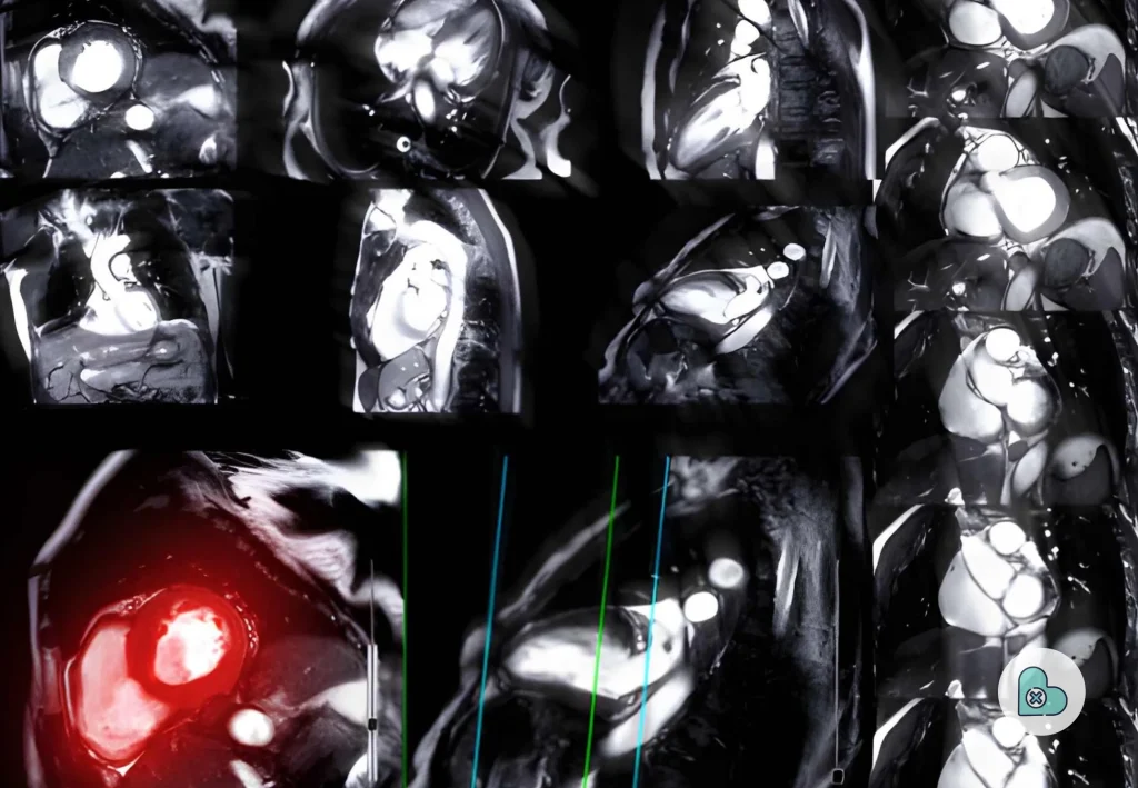
2. Introduction of 3D Imaging
To address the problems with flat images, scientists developed 3D echocardiography. This technology builds a three-dimensional model of the heart on a screen. It allows the doctor to rotate the image and look at the heart from every possible angle. This is a huge leap in cardiac imaging technology.
The benefits of 3D views include a clearer view of the heart’s structure. It improves the accuracy of every measurement the doctor takes. This leads to a more detailed diagnosis and less guesswork. When a doctor can see the heart in 3D, they feel much more confident in their treatment plan. It is a major part of modern healthcare technology.
3. The Breakthrough of 4D Ultrasound
The next massive step in this journey is a 4D ultrasound. If 3D gives you depth, 4D adds the element of time. This means 4D cardiac imaging shows the heart moving in high definition, in real time. You aren’t just looking at a 3D model; you are watching the heart function as it happens.
This allows the medical team to monitor the heartbeat in real time. They can study exactly how the valves move and spot tiny problems that 2D or 3D might miss. Real-time heart imaging is very helpful for complex surgeries. It provides a level of clarity that was simply impossible a few years ago. It is truly the gold standard of real-time ultrasound imaging.
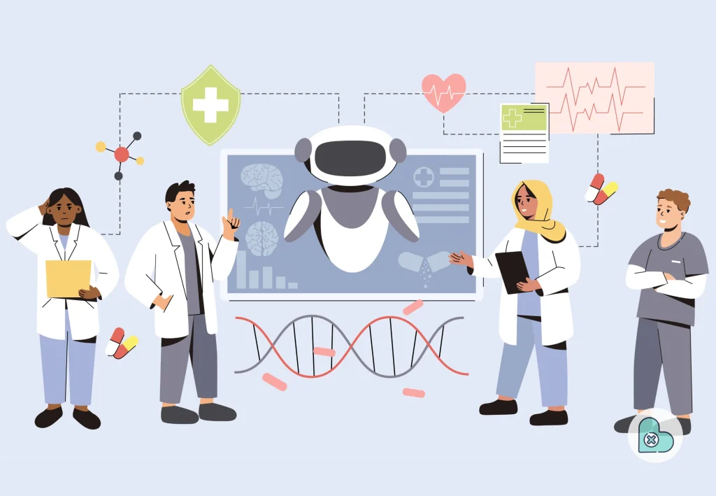
Key Technologies Driving the Future
_ freepik
AI in Cardiac Imaging
Artificial intelligence in healthcare is changing everything we know. In cardiology, AI in cardiac imaging helps automate boring tasks and makes every scan more accurate. AI can analyze thousands of images per second to detect tiny patterns. It acts like a second pair of eyes for the doctor.
AI-powered diagnostics can guide a technician to the optimal spot for the probe. This means even a less experienced worker can get a perfect image. This makes diagnostic imaging systems more reliable and much faster. It reduces the risk of human error and ensures that every patient receives the best possible care.
Machine Learning in Cardiology
Machine learning in cardiology is a subset of AI that gets smarter over time. It looks at millions of data points from different patients to predict risks. It helps detect heart disease early by spotting trends that humans might miss. This technology allows doctors to move from just reacting to problems to stopping them before they start.
Contrast-Enhanced Imaging
Sometimes, a regular cardiac ultrasound doesn’t show enough detail. That is where contrast-enhanced imaging comes in. Doctors use a safe liquid agent to make the blood show up clearly on the scan. This is great for blood flow imaging and finding blocked arteries. It provides a much more precise look at damaged heart tissue.
Point-of-Care Ultrasound (POCUS)
Portable devices are making waves in the medical world. POCUS ultrasound allows doctors to scan a patient at the patient’s bedside. These portable ultrasound devices are small enough to carry in a pocket. They provide point-of-care ultrasound with instant results. This is a lifesaver in emergency rooms where every second counts.
Telecardiology and Digital Solutions
Telecardiology connects specialists with patients who live far away. Doctors can share cardiovascular imaging files instantly over the internet. This enables remote cardiac diagnosis, saving patients from travelling long distances. It is one of the most important digital healthcare solutions for people living in rural areas.
How Advanced Imaging is Changing Healthcare

_ freepik
Advanced cardiac imaging is not just about shiny new toys. It is improving the whole hospital system. One of the biggest wins is a faster diagnosis. With smart imaging systems, you get results in minutes, not days. This allows the team to start life-saving treatments immediately.
It also leads to much better accuracy. Modern healthcare technology reduces mistakes by providing clear, sharp images. When the images are perfect, the cardiac treatment planning is perfect too. Doctors know exactly what to do before they ever step into an operating room. This reduces the need for “exploratory” surgeries and keeps the patient safe.
The patient experience is also much better. Most non-invasive diagnosis methods are painless and quick. Patients don’t have to deal with the stress of long waits or the fear of needles. This builds trust between the patient and the doctor. High cardiac imaging accuracy means the patient knows they are getting the right treatment the first time.
Real-World Applications of Cardiac Imaging

_ freepik
- Early Detection of Heart Disease: This is the most important use. Catching a heart issue early can double a patient’s chance of survival. Heart scan technology finds hidden problems before they cause a heart attack.
- Monitoring Long-Term Conditions: For people with chronic issues, heart disease monitoring is a part of daily life. Doctors use regular scans to see whether the medicine is working or whether the heart is getting stronger.
- Surgical Guidance: During heart surgery, doctors use real-time heart imaging to see where to place stents or valves. This improves precision and makes the surgery much safer for everyone involved.
- Emergency Care: In an ambulance or a trauma bay, portable ultrasound devices help doctors make split-second decisions. This is cardiology innovations at its finest—saving lives on the go.
Challenges in Cardiac Imaging
Even with all these medical imaging innovations, there are still some bumps in the road. The biggest issue is the high cost of the equipment. Advanced cardiac imaging systems can cost hundreds of thousands of dollars. This makes it hard for small clinics to get the latest tools. We need more affordable healthcare technology advancements in the Future.
Another challenge is the need for trained experts. A smart machine still needs a smart person to run it. Technicians need extensive training to use 4D ultrasound and AI tools correctly. Data management is also a challenge. High-quality scans create massive files. Storing these safely and sharing them between hospitals can be very complex.
The Role of 4D Ultrasound in the Future
As we look ahead, 4D ultrasound will become the heart of every clinic. It offers a live view of heart function analysis that no other tool can match. Because it is so clear, it helps in detecting even the smallest valve leaks. As technology becomes cheaper, we will see it in every doctor’s office, not just in big hospitals.
In the Future of healthcare imaging, 4D will likely be combined with virtual reality. Imagine a doctor putting on a headset to “walk through” a patient’s heart. This is no longer science fiction. It is the direction of cardiac imaging trends. This level of detail will make heart surgery safer than ever before.
Future Trends in Cardiac Imaging
The Future of cardiac imaging is all about being “smart.” We will see smart healthcare systems that talk to each other. Your cardiac health monitoring data from a watch might be sent directly to your doctor’s imaging software. This creates a connected loop of care that never stops.
Automation will also play a huge role. Many of the manual steps in a heart scan will be handled by AI-powered diagnostics. This reduces the workload for tired doctors and nurses. Most importantly, we are moving toward personalized care. Every cardiac treatment planning session will be based on the patient’s unique 4D heart model.
Finally, we will see better global accessibility. Thanks to portable ultrasound devices, a doctor in a remote village can get the same high-quality scan as a doctor in a big city. This is the ultimate goal of medical imaging innovations. We want to provide top-tier heart imaging techniques to every person on the planet, regardless of where they live.
Final Thoughts
Cardiac imaging has come a long way from the grainy black-and-white screens of the past. From the humble echocardiography to the mind-blowing 4D ultrasound, the progress is amazing. Today, we can see the heart in ways our grandparents never dreamed of. This leads to a better heart disease diagnosis and faster, more effective treatments.
The Future is very bright. Technology like AI in cardiac imaging will continue to grow and save lives. In the end, it is not just about the fancy machines or the fast computers. It is about the people. It is about giving every patient a chance at a long, healthy life. Cardiovascular imaging is the key to building a healthier future for everyone.


Leave a Reply